Our Advanced Diagnostic Technology
Optical Coherence Tomography
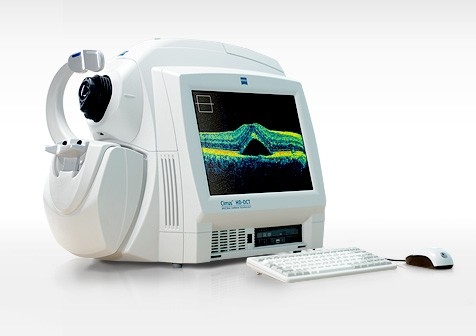
Optical coherence tomography, also known as OCT, is an imaging system that uses light waves to produce a high-resolution view of the cross-section of the retina and other structures in the interior of the eye. The images can help with the detection and treatment of serious eye conditions such as:
- Macular hole
- Macular swelling
- Optic nerve damage
- Age-related macular degeneration
- Macular pucker
- Glaucoma
- Cataracts
- Diabetic eye disease
- Vitreous hemorrhage
OCT uses technology that is similar to that of a CT scan of internal organs. With the scattering of light it can rapidly scan the eye to create an accurate cross-section. Each layer of the retina can be evaluated and measured and compared to normal, healthy images of the retina.
The OCT exam takes about 10 minutes to perform in your doctor’s office, and usually requires dilation of the pupils for the best results. We use the latest model, the Zeiss Cirrus 6000, which was released in late 2019.
Digital Fundus Photography and Angiography
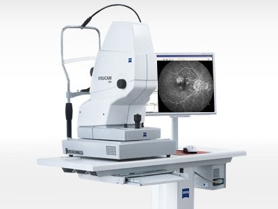
A fundus photograph is a specialized form of medical imaging. Using a customized camera with high-powered lenses that are mounted to a microscope, photographs are taken of the back of the eye by focusing light through the cornea, pupil and lens. Fundus photographs are used to identify or monitor a wide variety of ophthalmic conditions.
To begin the process, the pupil is dilated with eye drops. The patient will be asked to stare at a fixed device, keeping the eyes focused and still. There will be a series of flashes of light. The process usually takes no more than 10 minutes.
Some of the ophthalmic conditions fundus photography is used for including:
- Glaucoma
- Diabetic retinopathy
- Macular edema
- Microaneurysm
- Optic nerve
Digital fluorescein angiography is used to evaluate blood vessel flow in the retina, choroid, and optic nerve. It provides diagnostic information and helps to monitor the progress of treatment for many conditions in the eye. Intravenous contrast dye (similar to that used for cardiac angiograms) is injected into the patient’s arm. This is followed by a series of photographs as the contrast dye circulates in the eyes, giving the doctor valuable information about the condition being monitored or investigated.
Humphrey Visual Field
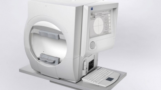
The Humphrey visual field is a diagnostic test to measure visual fields, or perimetry. The Humphrey visual field test measures the entire area of peripheral vision that can be seen while the eye is focused on a central point. During this test, lights of varying intensities appear in different parts of the visual field while the patient’s eye is focused on a certain spot. The perception of these lights is charted and then compared to results of a healthy eye at the same age of the patient to determine if any damage has occurred.
This procedure is performed in about 20 minutes and is effective in diagnosing and monitoring glaucoma. Patients with glaucoma will undergo this test on a regular basis to determine the progress of the disease. The Humphrey visual field test can also be used to detect conditions within the optic nerve of the eye, and certain neurological conditions as well.
Corneal Topography and Wave Front Analyzer
